Can a smartphone capture a 3D image of the optic nerve?
I asked myself that question and got to work.
As an initial step, I recorded an eye funduscopy (as «indirect monocular ophthalmoscopy»). This was done with a smartphone and a 20D magnifying glass.
After that, I captured the sharpest frames where the Optic Nerve (NO) could be clearly seen.
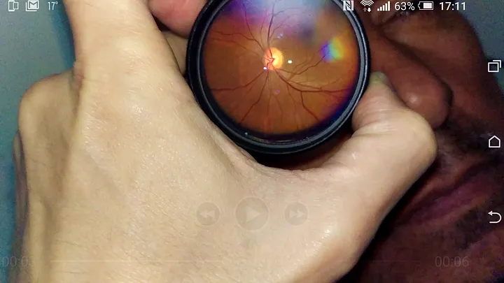
Then I picked the best.
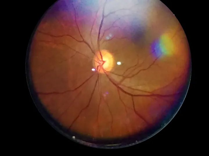
It is here that image processing begins to work its magic.
Through the use of a public domain program (ImageJ), I was able to extract different information from the image.
Those data were then represented differently until I reached the desired result.
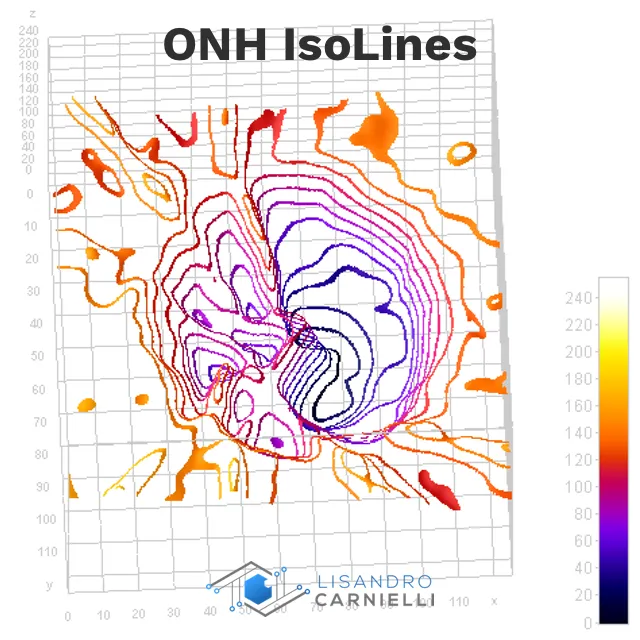
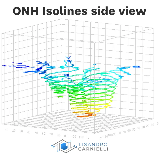
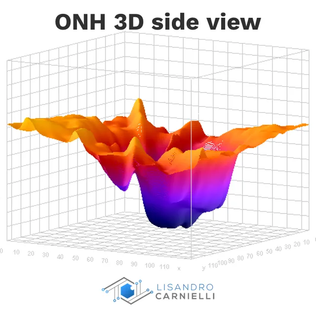
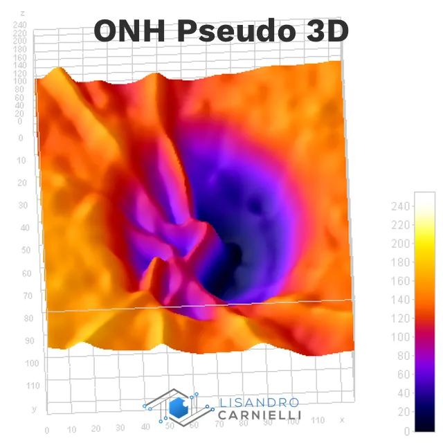
The final step was to compare the values of the image against a master image to which I previously knew the values of Disc Area, Disc Perimeter, Cup Area, and Cup/Disc Ratio (previously obtained by HRT-II).
To give to the patient, I generated a report based on these images and values.
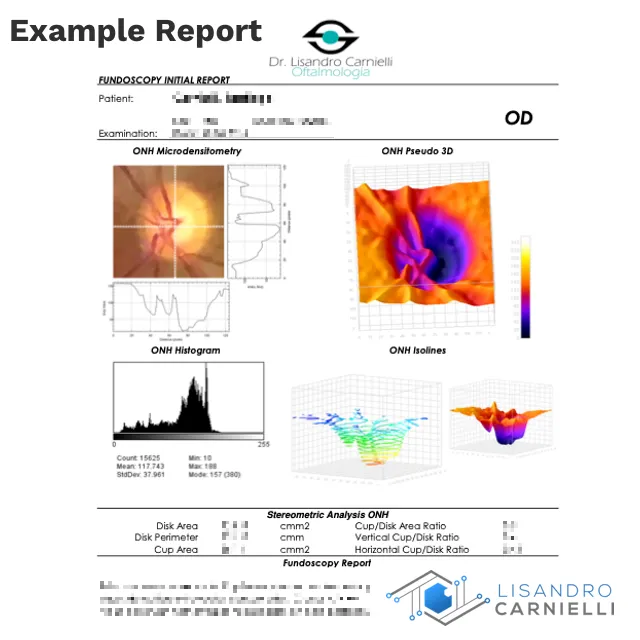
What do you think of the result?
- This is just an exercise, a proof of concept. I am aware that the 3D image is a pseudo-3D image and the values associated with cup depth depend on video color differences. No scientific validity is intended for the results obtained.

